Diagnostic technology for Ibuprofen-sucking, donut-squashing, claustrophobic patients and you.
Types of MRI
What’s the problem with most static radiology? It does not capture variable pressure points. Subsequently, viewers of spinal images lack the context of the amount of pressure that exerts pain. Lateral spinal images can be imaged while standing, lying straight, or with knees to chest. Even with knowledge of patient orientation, the area studied can have positional factors lost on static images.
There are at least two types of dynamic magnetic resonance imaging (MRIs). One refers to static images captured of a patient in a variety of positions. Scanning an extended neck significantly affects spinal cord compression severity compared with supine position. Comparing this alternate views can more specifically reveal the source of pain.
Conversely, a patient might remain stationary during multiple images while a concentrated contrast agent is injected. With a dynamic contrast-enhanced MRI, the contrast agent is measured as it passes through the brain or from blood vessels into extracellular space of tissues and back to the blood vessels. —NCBI
Dynamic Imaging Near You
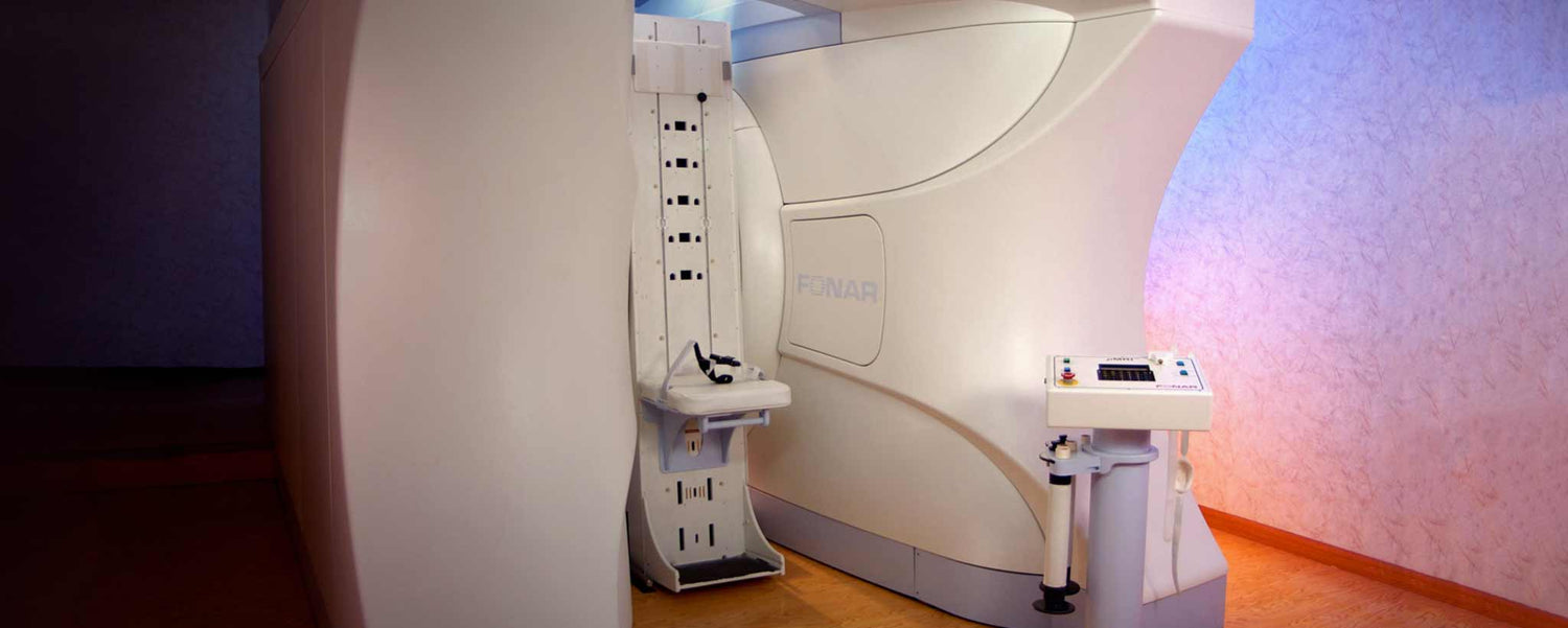
Not all radiology departments have dynamic MRI equipment. Because of the non-tubular design, facilities may reserve them for claustrophobic patients. Most scanners have a narrow tube through which a patient slides in and out on a conveyer belt. This does not provide much wiggle room for bending or sitting.
Manufacturers are offering innovative equipment that allow imaging in a variety of positions. Since most doctors rely on a specific radiology lab, patients may need to inquire about dynamic imaging outside the network.
If a photographer came back from the INDYCAR Grand Prix with only images of parked cars you would not get a sense of their speed. Similarly, images of parked bones do not always reveal extent of medical conditions.

Upright MRI machine in London.
It is important to visualize the weight-bearing coccyx when leaning backwards and forward. By means of dynamic images, doctors are able to diagnose coccydynia (coccalgia, coccygalgia, coccygial, coccygodynia) and sacroiliitis. This more accurately demonstrates mobile disc protrusion or hypermobile coccyx, often overlooked on presumption of pre-pubescent coccyx fusion.
Dynamic X-rays, or more often dynamic MRIs, are gaining diagnostic significance. A series of images allow the examiner to compare flexion vs. extension or standing vs. sitting. This means that during your next MRI, you do not need to feel like you are being sucked into a straw. Furthermore, the dynamic radiographs can be more informative to healthcare professionals. This leads to better treatment.
To support the writing of useful articles about technology, ClinicalPosters sells human anatomy charts, scientific posters, and other products online. You may sponsor specific articles, become a ClinicalNovellas Member, or remit a small donation.
ClinicalPosters sells human anatomy charts, scientific posters, and other products online to offset expense of the writing useful articles about technology. Slide extra posters into DeuPair Frames without removing from the wall.
Show your support by donating, shopping for ClinicalPins, becoming a ClinicalNovellas Member, or leaving an encouraging comment to keep the research going.
To support the writing of useful articles about technology, ClinicalPosters sells human anatomy charts, scientific posters, and other products online. You may sponsor specific articles or remit a small donation.
ClinicalPosters sells human anatomy charts, scientific posters, and other products online to offset expense of the writing useful articles about technology. Slide extra posters into DeuPair Frames without removing from the wall.
ClinicalPosters sells human anatomy charts, scientific posters, and other products online. You may remit a small donation or become a ClinicalNovellas Member.
You can support the writing of useful articles about technology by sponsoring specific articles, becoming a ClinicalNovellas Member, or remitting a small donation.


