Foot Skeleton with Ligaments and Muscles Model

$389

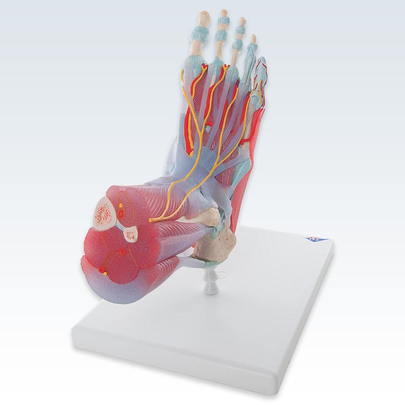
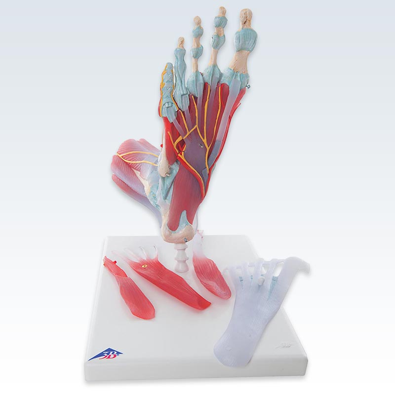
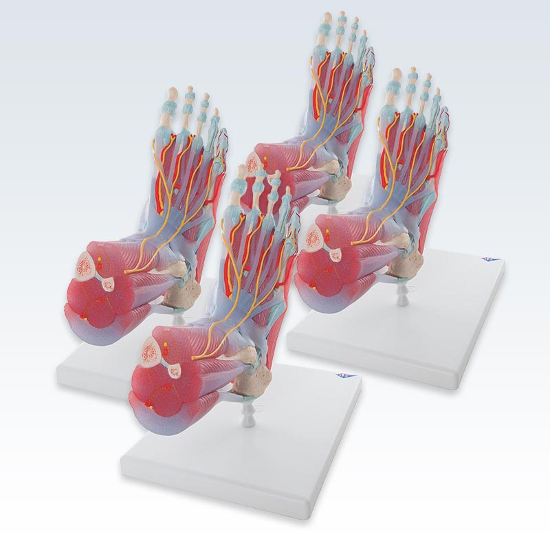
Exquisite detail of this 9.1 x 10.2 x 7.5 inch anatomical foot and lower leg model rexults in more compre­hen­sive patent consul­ta­tions. The model can be dis­assem­bled into 6 remov­able parts for deep study of the foot and ankle. The foot skeleton features not only the bones but also the muscles, tendons, liga­ments, nerves, arteries, and veins of the foot. [A]

Trust

The frontal view of the foot model features the extensor muscles of the lower leg. The tendons can be followed on their passage under the trans­verse and crucial crural ligaments all the way to their inser­tion points. In addition all tendon sheaths of the foot area are visible. On the dorsal portion of the foot the gastroc­nemius muscle is remov­able to reveal deeper anatomical elements.

The sole of the foot is repre­sented in three layers; the first layer displaying the flexor digitorum brevis. This muscle can be removed from the foot revealing the quadratus plantae, the tendon of the flexor digitorum longus, and the flexor hallucis muscle. This second layer is in turn remov­able to display even deeper anatomical details of the foot.

This model with ligaments and muscles is the best of its kind in quality and value. Purchase the best anatomy models for students, and doctors specializ­ing in orthopedic foot surgery and podiatry from Clinical­Posters, your anatomical model supplier.