


Female Pelvis with Ligaments, Vessels, Nerves, Organs Model
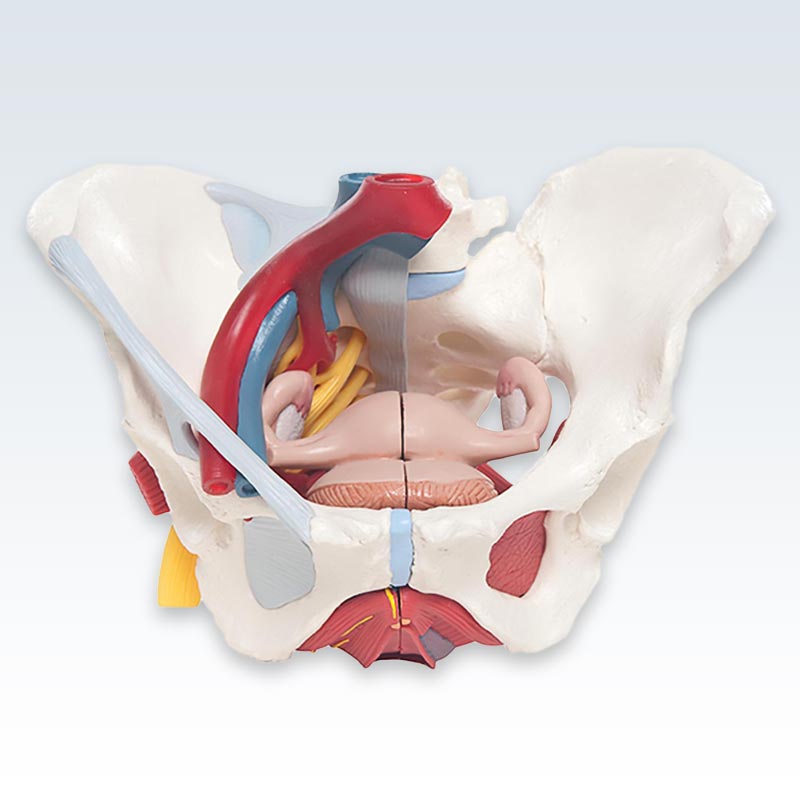
This life-size six-part 3B Scientific® anatomy model of a female pelvic region represents detailed information about the topography of bones, ligaments, vessels, nerves, pelvic floor muscles and female genital organs.
It presents the whole pelvic floor with partially removable mid-sagitally sectioned external anal sphincter, external urethral sphincter, deep and superficial transverse perineal and bulbospongiosus. Purchase the best anatomy models for students, and doctors specializing in orthopedic surgery, obstetrics and gynecology from ClinicalPosters, your anatomical model supplier. [A]
More Info
The rectum, uterus with fallopian tubes, ovaries and vagina are also removable and can be disassembled into both halves by midsagital section. The right pelvic half demonstrates the divisions and topographical anatomy of the common iliac artery, the external and internal artery and also of the common iliac vein and the external iliac vein. The right sacral plexus, right sciatic nerve and right pudendal nerve are also shown.
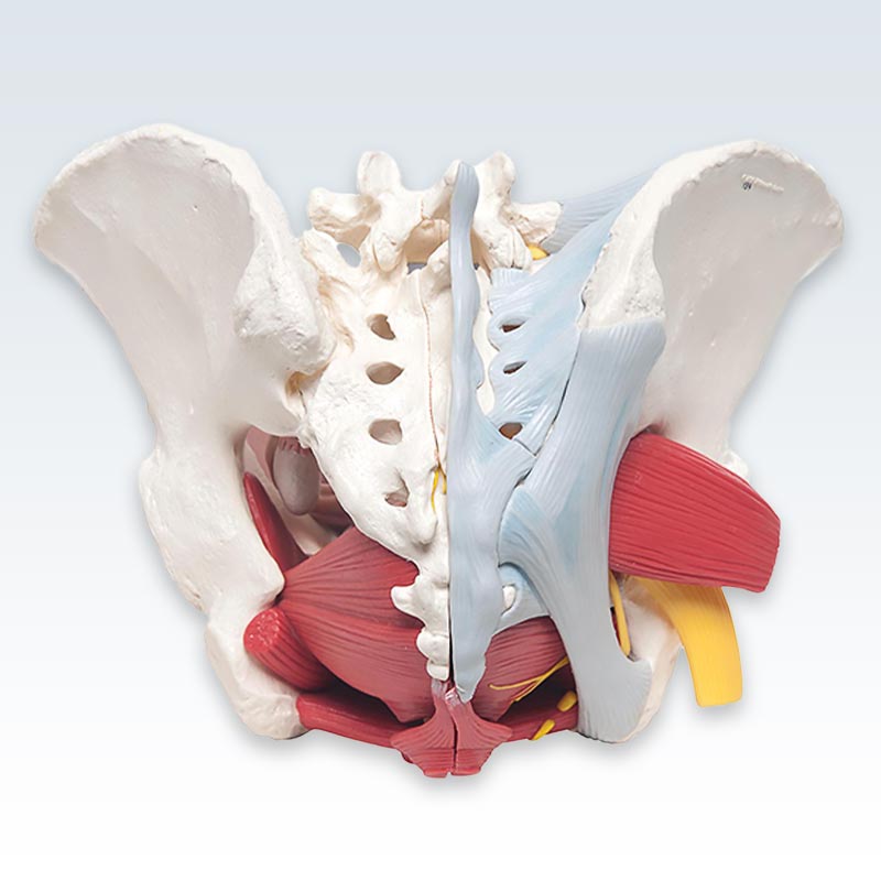
Bones and ligaments presented: Two hip bones, the pubic symphysis, the sacrum and the coccyx, the fifth lumbar vertebra with intervertebral disc. A midsagital section through the fifth lumbar vertebra, sacrum and coccyx, allow both halves of the pelvis to be disassembled revealing a part of the cauda equina in the vertebral canal. The left half of the fifth lumbar vertebral body is removable. The right half of the model shows the following pelvic ligaments: inguinal ligament, sacrotuberous ligament, sacrospinous ligament, anterior sacroiliac ligaments, iliolumbar ligament, anterior longitudinal ligament, interosseous sacroiliac ligament, posterior sacroiliac ligament and obturator.