

DermNet lists 130+ skin signs of systemic disease. Table expands diagnosis beyond acne or eczema and symptomatic cutaneous inflammation.
Which Skin Condition Concerns You?
Skin is a window to the soul. Common cutaneous disorders may be benign or could be an outward manifestation of internal or systemic disease. DermNet lists over 130 skin signs of systemic disease. This non-comprehensive table of 13 skin disorders expands diagnosis beyond acne or eczema and symptomatic cutaneous inflammation. [1–3]

It also reveals why some skin eruptions are resistant to topical dermatology treatments; the underlying condition must be addressed. Fingernail irregularities can also signal underlying disorders. [4]
| Cutaneous Disorder | Description | Internal Differential |
|---|---|---|
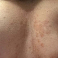
| Seborrheic Dermatitis | Chronic superficial inflammatory disease of the scalp, face (i.e. eyebrows and nasolabial folds), ears and central chest. | Possible association with systemic disease such as Parkinson’s disease and human immunodeficiency virus (HIV) infection or cerebrovascular accident. |
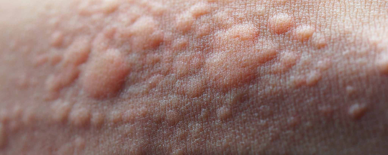
| Erythema Multiforme | Cutaneous hypersensitivity reaction. Macules, papules, plaques, vesicles, or bullae, often with a targetoid or iris appearance, often with distribution throughout extremities. | Usually caused by infection (herpes simplex virus or Mycoplasma pneumoniae) and less commonly by drug sensitivity. |
| Cutaneous Metastases | Metastases to the skin are usually flesh-colored to violaceous nodules that appear in close proximity to the primary neoplasm; most common sites are the scalp, neck, and trunk. | Cancers of the breast and large intestine are the most likely primary tumors to metastasize to the skin in women. |
| Paget’s Disease | Unilateral eczematous plaque of the nipple and areola. Persistent, eczematous plaque of the anogenital or axillary regions. | Strongly associated with an underlying invasive carcinoma of the affected breast or ductal carcinoma in situ. |
| Acanthosis Nigricans | Smooth, velvet-like, hyperkeratotic plaques in intertriginous areas. | Type I is associated with malignancy. Type II has no malignancy association. Type III acanthosis nigricans is associated with obesity and insulin resistance. |
| Cowden’s Syndrome | Wartlike growths around the mouth, nose, and ears. | Cancer of the breast, endometrium, or thyroid gland; and thyroid disease (adenomas or goiter), mental retardation, and fibrocystic disease of the breast. |
| Sweet’s Syndrome | Painful erythematous to violaceous plaques on the face, extremities, and trunk. Most have a fever, malaise, arthralgias, myalgias, and conjunctivitis. | Inflammatory bowel disease, bowel bypass syndrome, and pregnancy. It occasionally occurs as a reaction to certain medications. |
| Amyloidosis | Papules on the eyelids and extremities that become purpuric and ecchymotic due to increased blood vessel fragility secondary to amyloid infiltration of the vessels. | May be a sign of multiple myeloma. |
| Paraneoplastic Pemphigus | Intractable stomatitis and blisters on the trunk and extremities. | Strong association with non-Hodgkin’s lymphoma, chronic lymphocytic leukemia, and Castleman’s disease with or without myasthenia gravis. |
| Sarcoidosis | Red to purple indurated plaques of the nose (lupus pernio), midfacial papules, annular plaques, and plaques or nodules on the trunk and extremities. | Multisystem, granulomatous disease of the lungs, bones, central nervous system, lymph nodes, eyes, and skin. |
| Erythema Gyratum Repens | Red to purple indurated plaques of the nose (lupus pernio), midfacial papules, annular plaques, and plaques or nodules on the trunk and extremities. | Strong association with lung cancer; association with breast, cervical and gastrointestinal cancers. |
| Psoriatic Arthritis | Silvery, itchy scales and painful red patches can appear on the scalp, eyelids, ears, mouth and lips, skin folds, hands and feet, nails, or other body areas optionally present with asymmetric fusiform swelling of the distal interphalangeal joints, in association with oligoarthritis and tenosynovitis. | Can resemble rheumatoid arthritis. |
| Dermatomyositis | Scaly, telangiectatic plaques with atrophy and hypopigmentation on the face, neck, trunk, and extremities; malar erythema; and nail abnormalities (periungual telangiectases and cuticular hypertrophy). | Symmetric proximal muscle weakness (myositis); photosensitivity; papules and plaques on the hands, elbows, and knees. |
Sweet Reaction
A patient noticed the sudden onset of painful, erythematous lesions on her forehead. It appeared to be an insect bite, vasculitis, atypical pyoderma gangrenosum, Behcet’s disease, or possibly a herpes virus infection. But this turned out to be more than a dermatological concern.
The patient had acute febrile neutrophilic dermatosis, first described by Dr. Robert Douglas Sweet in 1964. [5] Sweet’s syndrome may also be a reaction to a more serious condition, such as leukemia, or a response to certain medications. [6] Protecting your skin from prolonged sun exposure is a good way to avoid recurrences of Sweet’s syndrome.
A person cannot easily see within the body without the aid of special equipment. So when something is going wrong inside, the skin on the outside may provide clues.
Included within the 19 illustrations, Anomalous Areolae depicts dermatitis, nevoid hyperkeratosis, Paget’s disease, carcinoma and malignant cancer of the breasts.
Graphic photos: Skin conditions
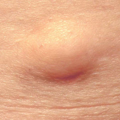 Lipoma | 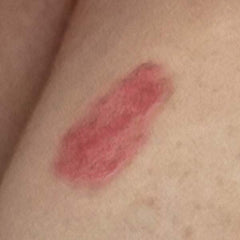 Rash | 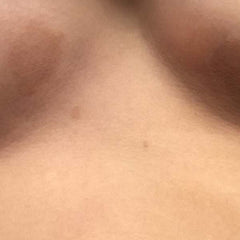 Fungus | 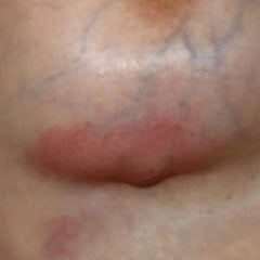 Cyst |
 Fungus | 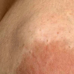 Tumor | 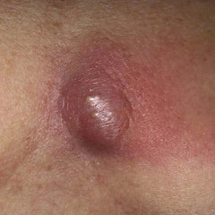 Abscess | 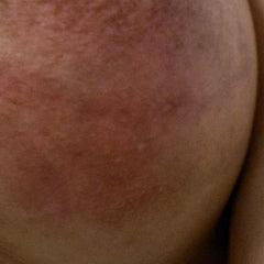 Dermatitis |
A breast abscess is a localized collection of pus within the breast tissue, often preceded by mastitis. About 3 percent of women who experience breast inflammation will develop an abscess.
Skin and Common Disorders is an anatomical poster with 23 individual illustrations that highlight 22 of the most common lesions seen by dermatologists. Types of skin lesions are described: fissure, ulcer, cyst, macule, papule, wheal, vesicle, pustule, bulla, and nodule. It also shows and describes acne (closed and open comedo), actinic keratosis (solar keratosis), junctional nevus (mole), basal cell carcinoma, squamous cell carcinoma, verruca vulgaris (wart), seborrheic keratosis, dermatofibroma and urticaria (hives). Order a poster to hang on your wall for reference.


 Romance & Health Intertwine. Fall in love with a captivating romance miniseries that explores the essence of well-being. Become a ClinicalNovellas library member for heartwarming tales.


To support the writing of useful articles about dermatology, ClinicalPosters sells human anatomy charts, scientific posters, and other products online. You may sponsor specific articles or remit a small donation.
ClinicalPosters sells human anatomy charts, scientific posters, and other products online to offset expense of the writing useful articles about dermatology. Slide extra posters into DeuPair Frames without removing from the wall.
Show your support by donating, shopping for ClinicalPins, or leaving an encouraging comment to keep the research going.
To support the writing of useful articles about dermatology, ClinicalPosters sells human anatomy charts, scientific posters, and other products online. You may sponsor specific articles or remit a small donation.
ClinicalPosters sells human anatomy charts, scientific posters, and other products online to offset expense of the writing useful articles about dermatology. Slide extra posters into DeuPair Frames without removing from the wall.
ClinicalPosters sells human anatomy charts, scientific posters, and other products online. You may remit a small donation.
You can support the writing of useful articles about dermatology by sponsoring specific articles or remitting a small donation. Visible content is optimized for device size.

